Archive for February, 2013

Role of Primitives, Patterns, Weights, and Modules during Locomotor Development

February 26th, 2013

Primitives. Hey! What do you mean by “Primitives”?!

According to Giszter et al. (2010), the “term ‘primitive’ may best indicate the idea of a set of building blocks or developmental bootstrap elements that is used in a constructive or compositional fashion” from which “larger modules can be made.”

Thus, these primitive building blocks can be hypothesized to define modules of muscle activations that are a combination of basic patterns and weights distributed to the muscles composing the module (see Lacquaniti et al. 2012).

Dominici etal 2012 F1

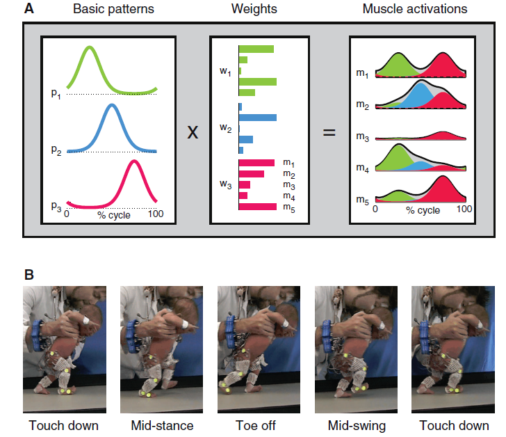
Fig. 1. (A) Schematic of motor modules (m). Simulated example of muscle activity profiles as weighted (w) sum of basic patterns (p): mi(t)=Σjpj(t)wij. The outputs of the first (green), second (blue), and third (magenta) modules are summed together (i = 5, j =3) to generate overall muscle activation (black envelope). (B) Illustration of a step cycle in a 3-day-old newborn.

Click to keep reading this post!

NOPE; Spinal Interneurons are NOT Active In A Graded Manner

February 19th, 2013

Are the motoneurons like those involved in swimming, for example, by a zebra fish, recruited in a graded manner? Yup

Then, are the active set of spinal interneurons graded during increases in swimming speed? NOPE!!!!

A fascinating study by Fetcho, McLean, and colleagues (2008), entitled “Continuous Shifts in the Active Set of Spinal Interneurons during Changes in Locomotor Speed” found these finding in the larval zebra fish. Instead of repeating what they wrote, here are some intriguing figures:

Figure 1, Parts a – d. Gradation of movement. These parts of Figure 1 show slow swimming in a; movement around vertical axis (i.e., yaw) for head, midbody, and tail in b; yaw for evoked swimming in c; and yaw for spontaneous swimming in d. Parts c and d demonstrate that “fish can smoothly grade between the fastest and the slowest swimming movements without any obvious discontinuity in the axial bending pattern.”

McLean etal Cont shifts 2008 Fig 1 top1

Figure 1 a-d.  Analysis of real and fictive evoked swimming movements. (a) Consecutive overlapping images of a bout of swimming elicited by a tactile stimulus to the tail (at asterisk). A frame extracted from the montage (gray arrows) shows the regions selected for kinematic analysis at the head (H), midbody (M) and tail (T). Images were captured at 1,000 Hz (images 1–9, every 4 ms; 10–12, 8 ms; 13–14, 16 ms; 15–18, 32 ms). (b) Automated analysis of yaw at three points along the body, from the bout in a. Only the tail showed any noticeable movement at the end of the bout, when swimming was slowest. (c) Plots of head and tail yaw from 12 evoked swimming bouts in 12 larvae. The degree of head and tail yaw decreased as a function of swimming frequency. Open circles are raw data points, whereas closed circles represent means (± s.d.) from data binned at 5-Hz intervals (for example, 15–20, 20–25, etc.). (d) A similar plot for spontaneous bouts of swimming (12 from the same 12 larvae), whose values are comparable to the lower end of evoked swimming frequencies. Only the first episode of the five analyzed in each fish is shown in c and d.

 
Keep reading this poat.

What Is the Primal Movement Starter?

February 12th, 2013

ooh! OOH! ESCAPE!

There is no doubt that escaping capture was critical to survival.

So, what starts escape and also other forms of movement? Let’s start by looking at a primitive animal: the leech.

Escape for the leech is swimming, as long as it is in relatively deep water. A tap on its tail will result in fast swimming!

How does a leech swim? It creates an approximately sinusoidal, undulatory traveling wave. Contraction and relaxation of dorsal and ventral longitudinal muscles are primarily responsible for swimming undulations (Lamb & Calabrese, 2011).

NOTE: for scientists, there is neural activity ‘sign’ that an isolated or nearly isolated leech preparations would be swimming if it was in an intact leech. The ‘sign’ is a bursting pattern of a dorsal excitatory motoneuron in these preparations, and the behavior is called “fictive” swimming. Researchers frequently refer to “fictive swimming” as a “swim”, “swim episode”, “swimming”, and “swim activity” (Mullins et al. 2011a).

What are the pathways activated in these muscles? At the base is a central pattern generator (CPG) circuit that is composed of complex segmental oscillators which rely heavily on intersegmental connectivity (Lamb & Calabrese, 2011).

Midbody ganglia contain a bilateral, triphasic oscillator CPG circuit composed predominately of bilaterally paired interneurons. And the output from these oscillator interneurons controls the activity of the excitatory and inhibitory motoneurons. It is these interneurons that provide the final common pathway to the longitudinal muscles used for swimming. Otherwise, a leech is simply resting.
Keep reading this poat.

Beginning Objectives for 2013

February 6th, 2013

Here is what is planned for this website for the beginning of 2013:

First, this is the list of next blog Posts:

  1. What is/are the Primal locomotor initiator(s)? And where is it/are they located?
  2. Does the recruitment of spinal interneurons increase according to the motorneuron cell size principle?
  3. How are the recruitment of muscle patterns, i.e., muscle synergies, involved for postural changes during locomotion? Is the midbrain reticular formation or basal ganglia directing any change in motor pattern selection?
  4. What senses muscle force exerted during locomotion? What senses pain? What senses exertion especially during high intensity sport?
  5. How does the estimation of the sense of force, pain, exertion, and effort become reported?  And how is exertion sense used in producing a specific work rate?
  6. What increases the rating of perceived exertion (RPE) during a constant work rate? Is the mechanism related to the Central Governor Model?

Second, the increase in RPE(Delta RPE) will be explored as a Critical Limiting Factor for prolonged endurance. That is, is Delta RPE a Critical Limiting Factor? Or, what are associated with Delta RPE that are Critical Limiting Factors? Also, this means an updating of the background Pages about Limiting Mechanisms, and in the process of updating, perhaps writing a Review Page about Delta RPE and Critical Limiting Factors.

Third, preparations will be made for the next series of blog Posts exploring the mechanisms for pain and fatigue.

Fourth and last, steps will be taken for improving the sharing of this website’s Pages and Posts.