Student: The Airways are connected to the Pulmonary Stretch Receptors that are connected to the Vagal Nerves. Then they are connected to what next?
Professor: Before we get to the your intriguing question, let’s consider all the ‘inputs’:
- Airway smooth muscle
- Diaphragm skeletal muscle
- Chest wall skeletal muscle
- Lung airways and tissue
Shown below are two models for dyspnea; the left is Davenport (2007) and the right is Weiser, Mahler, et al. (1993). The bottom of BOTH diagrams displays the lung inputs that can become the signals for uncomfortable breathing:
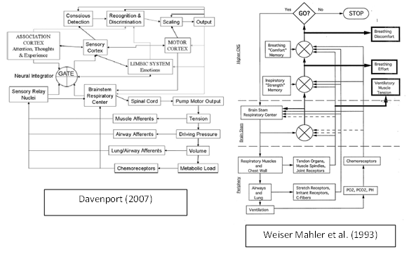
Student: BOTH models’ bottom loops are almost IDENTICAL.
Professor: And in another review, Davenport & Vovk (2009) show a similar diagram, which we will see shortly. And in it, they show an earlier structural model that is hierarchical and is divided into three levels:
- Cortical Processing,
- Sub-cortical Central Nervous Processing, and
- Sensory Signal Transduction.
Professor: these loops are within the bottom Sensory Signal Transduction level. The common elements for both models above are the:
- Brainstem Respiratory Center,
- Respiratory Pump Outputs and their actions,
- Sensory Receptor Afferents, and
- Sensory Feedback to the Brainstem Respiratory Center.
Student: Davenport (2007) also identifies Sensory Relay Nuclei.
Professor: On our 1993 model we show a collateral from Brain Stem Respiratory Center that is sending an Efference Copy to a comparator in brainstem. Any difference between the Efference Copy and the Pulmonary Afferents Feedback becomes an Error Signal.
Student: Aha, now you are answering my question about, “Where the signals go after arriving in brainstem?”
Professor: The Answer is:
- The vagal fibers synapse in the brainstem perhaps directly on the Brainstem Respiratory Center.
- The second-order neurons are part of Sub-Cortical Central Nervous Processing.
Professor: In a nutshell, the structures at this level, at least include the Cingulate , Hypothalamus, Hippocampus, Amygdala, Thalamus, and Caudate as part of the Basal Ganglia, as shown in the hierarchy below:
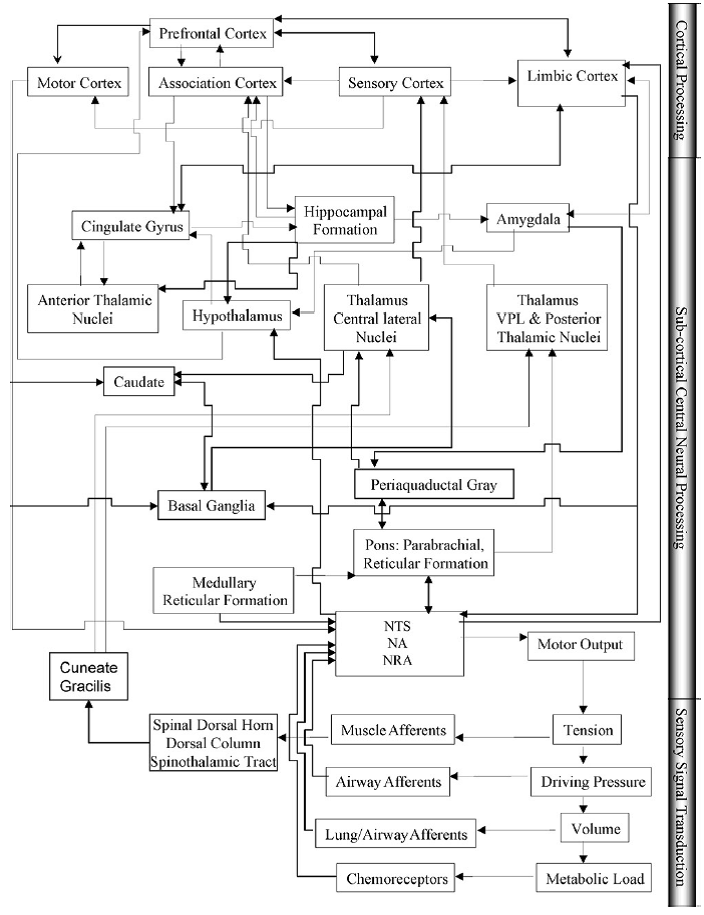
Fig. 3. Model of cortical and subcortical brain areas activated by stimuli that elicit dyspnea. The interconnections are based on anatomical relationships between the brain areas. See text for detailed descriptions. Abbreviations: NTS, nucleus of the tractus solitarius; NA, nucleus ambigulais; NRA, nucleus retroambigualis. [From Davenport & Vovk (2009)]
Professor: I would add the Insula to these structures, say above right Thalamus VPL & Posterior Thalamic Nuclei box and below the Amygdala box.
Student: You keep on telling us to disregard structures per se and more importantly look at regional functional connectivity, i.e., to concentrate on the networks that utilize various structures.
Professor: In that case, we are now talking about the core network: the Limbic Network
Student: Which parts of the Limbic Network extend into, or perhaps out of, the Sub-cortical Central Nervous Processing level?
Professor: According to Lisa Feldman Barrett and her group (first see Oosterwijk et al. (2012), this is the Limbic Network:
| ‘limbic’ [27] | medial temporal lobe, subgenual anterior cingulate cortex, medial, and lateral orbitofrontal cortex* |
|
core affect generation: representing visceromotor states from prior experience or engaging visceromotor control of the body to create the core affective tone (pleasure or displeasure with some degree of arousal) that is a basic feature of all conscious experience and that directs basic approach/withdrawal behaviors. |
*” Although Yeo et al. [27] did not include subcortical structures in their analysis,
- “We include subcortical structures in this network based on their known anatomical connections.
- “We include the nuclei of the basal ganglia, which are involved in orchestrating effortful behavior and motor control [81].
- “We also hypothesize that the central nucleus of the amygdala, which is involved in producing autonomic responses [81], and the midbrain periacqueductal gray, which is involved in coordinating coherent physiological and behavioral responses [81], are part of this network.
- “The basal ganglia, the amygdala, and the periacqueductal gray all project to the ventromedial prefrontal cortex (vmPFC), the major cortical site in Yeo et al.’s limbic network.” [Emphases and reformatting added. For the references cited, see the Lindquist KA, Feldman Barrett L. (2012) review.]
Professor: In regard to uncomfortable breathing, we can trace neural activity from receptors along sensory afferents to the spinal cord. Then some neural information can be sent to the Brainstem Respiratory Center, and also it is fed into the Limbic Network via the Thalamus
Student: I would like to learn more about these networks!
Professor: How about analyzing distributed neural networks used by meditators while noticing their mind wandering and switching their mental activities back to their breathing?
Student: Okay, I’m game.
Take Home Message: Uncomfortable breathing is reported as a mismatching of signals from ventilatory drive or airway with the resulting ventilation or discomfort memory,
Next: Awareness occurs from increased activity of Salience Network to inhibit Default Mode Network activity resulting In a wandering mind.
References:
Davenport PW (2007) Chemical and mechanical loads: What have we learned? In: O’Donnell DE, Banzett RB, Carrieri-Kohlman V, Casaburi R, Davenport PW, Gandevia SC, Gelb AF, Mahler DA, Webb KA. (2007) Pathophysiology of Dyspnea in Chronic Obstructive Pulmonary Disease: A Roundtable. Proc Am Thorac Soc 4: 147–149
Davenport FW, Vovk A. (2009) Cortical and subcortical central neural pathways in respiratory sensations. Resp Physiol Neurobiol 167: 72–86.
Lindquist KA, Feldman Barrett L. (2012) A functional architecture of the human brain: emerging insights from the science of emotion. Trends Cogn Sci 16: 533-540.
Oosterwijk S, Lindquist KA, Anderson E, Dautoff R, Moriguchi Y, Feldman Barrett L. (2012) States of mind: emotions, body feelings, and thoughts share distributed neural networks. Neuroimage 62, 2110–2128.