Student: Today my asthma lots of wheezing, and I was really terrible at meditating, at focusing on my breath.
Professor: You were focused on wheezy breathing, eh? That must have been very distracting.
Student: Yeah. I kept on thinking of using my ‘PUFFER’ instead of ‘THE BREATH’.
Professor: Sorta sounds like:
- “Mind wandering and attention during focused meditation”,
- That’s the title of an intriguing article by Hasenkamp et al. (2012).
Professor: They analyzed distributed neural networks used by meditators who would notice their mind wandering and then switch their mental activities back to theirbreathing. Shall we look into their main results?
Student: Yes, let’s do that.
Professor: Figure 1 for Hasenkamp et al. (2012) is shown below. Their panel A, your “Thinking about Wheezing” could be an example of what they called “Mind-wandering” (MW). When a participant detected MW, he or she pressed a button. The 3 s containing the button press defined the AWARE phase. The prior 3 s was MW baseline phase. And the next 3 s was the phase that the meditators made a SHIFT redirecting their attention back to focusing on THE BREATH. Then the next 6 s was defined as the sustained FOCUS phase (see below):
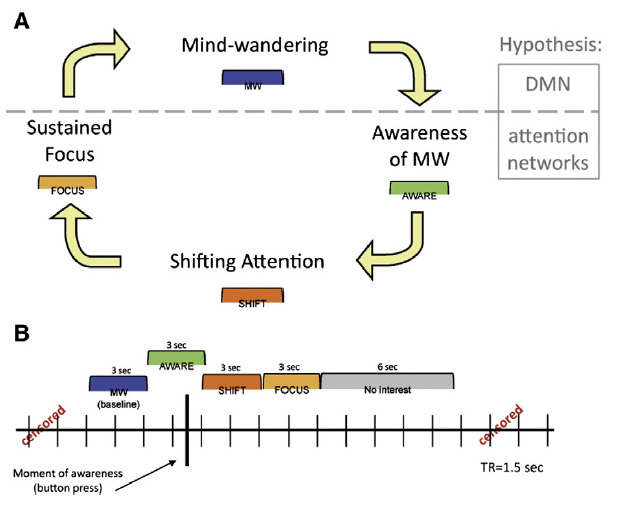
Professor: Above, Panel B of their Figure 1 shows their
“analytical model for construction of phases surrounding each button press (represented by the heavy black vertical line). While the button press is represented here in the middle of a TR [Time of Repetition for the MRI scanner], note that the timing of the button press within a TR will be variable.”
Student: Well, what areas were active during the AWARE phase?
Saliency Network
Professor: The AWARE areas are many of those Oosterwijk et al. (2012) and others call the “Saliency Network”. Its functions are thought to provide, “Body-Directed Attention”:
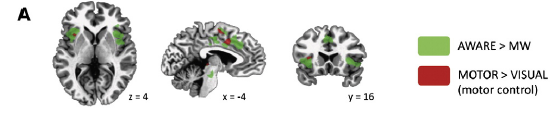
Professor: Above, In Panel A of their Figure 2, Hasenkamp et al. state that, “Activations during the AWARE phase are in green…. Voxels that were also significantly active during the motor control task (MOTOR>VISUAL [baseline]; p<0.005) are shown in red. Prominent activity was detected in dorsal ACC [Anterior Cinculate Cortex] and frontoinsular cortex.” [Emphases added.]
Professor: in their Table 2, they list the Brodmann areas that were more active in the AWARE phase compared to the WM baseline phase:
| AWARE > MW | Brodmann area |
| L pre/postcentral gyrus | 1,2,3,4 |
| L. posterior insula | 13 |
| R anterior/middle insula | 13,47 |
| Dorsal ACC | 24,32 |
| L anterior/middle insula | 13,47 |
| Midbrain | – |
| L superior parietal | 7 |
| L SFG/MFG | 10 |
Abbrev: ACC: anterior cingulate cortex, MFG: middle frontal gyrus, SFG: superior frontal gyrus.
Student: How well do these areas match the saliency network as defined by Oosterwijk et al. (2012)?
Professor: Well, below is the snippet from Table 1 for Oosterwijk et al. (2012):
| Network | Brain regions | Task domains | Psychological description and hypotheses |
| “salience network” (Seeley et al., 2007)or “ventral attention network”(Yeo et al., 2011; Corbetta & Shulman, 2002)or “cingulo-opercular network”(Vincent et al., 2008) | bilateral anterior midcingulate cortex (aMCC), anterior insula (AI) and mid-insula, frontal operculum, and parts of the pars opercularis and temporoparietal junction |
• cognitive control (Cole & Schneider, 2007)• stimulus-driven control of attention (Corbetta & Shulman, 2002)• set maintenance (Dosenbach et al., 2006)• maintaining sub-goals (Fincham et al., 2002)• anxiety (Seeley et al., 2007)• representation of the body (Craig, 2009)• pain (Lamm et al., 2010) | Body-Directed Attention: using representations from the body to guide attention and behavior. This ingredient might use changes in the homeostatic state of the body to signal salient events in the environment and regulate behavioral responses.
Hypothesis: Body feeling and Emotion > Thought |
Professor: Also, the bilateral anterior/mid-insula and anterior cingulate cortex are parts of Oosterwijk et al. (2012)’s salience network. Possibly the other AWARE areas, like pre/post central gyrus, superior and middle frontal gyrus, and midbrain, may be past of motor planning/selection prior to and during the button press.
Student: Also in the right column cell for Oosterwijk et al., they state, “Body-Directed Attention” is “using representations from the body to guide attention and behavior. This ingredient might use changes in the homeostatic state of the body to signal salient events in the environment and regulate behavioral responses.”
Professor: Hasenkamp et al. (2012) that these areas have been “implicated in a diverse range of cognitive processes, including:
- conflict monitoring and error detection,
- interoceptive- autonomic arousal,
- the moment of perceptual recognition,
- self-regulation,
- emotional aspects of pain,
- empathy,
- musical chills,
- pleasurable touch, and
- present moment awareness (reviewed in Craig, 2009; Seeley et al., 2007; Singer et al., 2009).
Student: In their experiment, that seems to mean detecting conflict, not on THE BREATH, and self-regulating to get back on THE BREATH. Yes, I will just notice my wheezing and keep my focus on THE BREATH.
Executive Attention Network
Professor: Now let’s look at the areas active during the SHIFT phase. A hint: many of them are from the Frontoparietal Network (or Executive Control network). Its functions are thought to provide: “Executive Attention”:
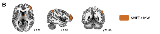
Professor: Above, in Panel B of their Figure 2, Hasenkamp et al. state “Activations in lateral PFC and posterior parietal regions [were found] during the SHIFT phase.”
Professor: In their Table 2, they list the Brodmann areas that were more active in the SHIFT phase compared to the WM baseline phase:
| SHIFT > MW | Brodmann area |
| R dlPFC/SFG/MFG/IFG | 8,9,10,46 |
| L caudate body/thalamus | – |
| R caudate body/thalamus | – |
| R inferior parietal | 40 |
| L inferior parietal | 40 |
| R MFG | 9 |
Abbrev: dlPFC: dorsolateral prefrontal cortex, IFG: Inferior frontal gyrus, MFG: middle frontal gyrus, SFG: superior frontal gyrus.
Student: How do these areas match the executive control network defined by Oosterwijk et al. (2012)?
Professor: Below is the snippet from Table 1 of Oosterwijk et al. (2012) for this network:
| Network | Brain regions | Task domains | Psychological description and hypotheses |
| “frontoparietal network” (Dosenbach et al., 2008; Vincent et al., 2008; Yeo et al., 2011)or “executive control network”(Seeley et al., 2007) | bilateral dorsolateral prefrontal cortex (dlPFC), inferior parietal lobe,inferior parietal sulcus, andaspects of the middle cingulate cortex (mCC) |
• task-switching (Crone et al., 2006)• alerting to a stimulus after a cue (Fan et al., 2005)• planning (Fincham et al., 2002)• rule-specific processing (Sakai & Passingham, 2006)• working memory (Sakai & Passingham, 2003) | Executive Attention: modulating activity in other ingredients to create a unified conscious field during the construction of a mental state (e.g., selecting some conceptual content when meaning is made of sensations and inhibiting other content; selecting some sensations for conscious awareness and inhibiting others).No specific hypothesis formulated |
Student: And in the right column cell for Oosterwijk et al., they state, “Executive Attention” is “modulating activity in other ingredients to create a unified conscious field during the construction of a mental state (e.g., selecting some conceptual content when meaning is made of sensations and inhibiting other content; selecting some sensations for conscious awareness and inhibiting others).
Professor: Hasenkamp et al. (2012) that these executive control network:
- “Acts on relevant stimuli (which are thought to be identified by the salience network) by re-orienting or directing attention while maintaining a goal (Corbetta and Shulman, 2002; Corbetta et al., 2008; Seeley et al., 2007).
- “Parietal elements of this network have been implicated specifically in attentional disengagement (Posner and Petersen, 1990), a process often accompanying re-orienting and also likely occurring during this phase.”
Student: Thus, what is known about the function of this network corresponds well with the hypothesized cognitive processing occurring in this phase: shifting or re-orienting attention from mind wandering back to the breath.
Working Memory
Professor: Interesting! The area active during the FOCUS phase is from the Executive Control network indicating more ” Executive Attention” happening:
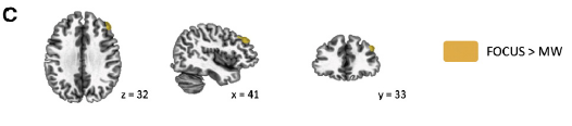
Professor: Above, in Panel C of their Figure 2, Hasenkamp et al. state: “Activation in dorsolateral PFC {was found} during the FOCUS phase.”
Student: Yes! This is one of the areas of the executive control network listed by Oosterwijk et al. (2012).
Professor: In their Table 2, they list the Brodmann areas that were more active in the FOCUS phase compared to the WM baseline phase:
| FOCUS > MW | Brodmann area |
| R dIPFC/MFG | 9 |
Abbrev: dlPFC: dorsolateral prefrontal cortex, MFG: middle frontal gyrus
Professor: Yes. What could be a function for this region?
Student: I would pick the following functions from the snippet for the executive control network in Table 1 for Oosterwijk et al. (2012):
- Selecting some sensations [better yet: actions] for conscious awareness and
- inhibiting others
Professor: This cluster of activity in the executive control network continued to be active from the SHIFT phase. The investigators, therefore suggested that:
- “This may represent persistent neural activity underlying working memory, or keeping the goal in mind, to maintain sustained attention on the focal object….
- The dorsolateral PFC has been specifically implicated in active rehearsal, which consists of “the repetitive selection of relevant representations or recurrent direction of attention to those items” (D’Esposito, 2007).
- “Active rehearsal would be central to the sustained attention we hypothesize is occurring in the FOCUS phase, providing repetitive selection of, or attention to, the breath.
- “The lack of activation in parietal elements of the executive network during this phase may be related to the role of the parietal cortex in disengagement of attention rather than in focusing.”
Default Mode Network
Professor: Finally, let’s look at the areas active during the baseline phase. Many of the areas are from the Default Mode Network (or Ventral Attention Network). Its functions are thought to provide: ” Conceptualization “:
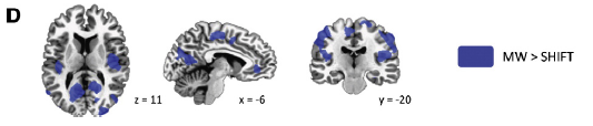
Professor: Above, in Panel D of their Figure 2, Hasenkamp et al. state: “Activations during MW phase included elements of DMN, as well as sensory and motor cortices and posterior insula.”
Professor: In their Table 2, they list the Brodmann areas that were more active in the WM baseline phase rather than in the following SHIFT phase:
| MW > SHIFT | Brodmann area |
| R posterior insula, | 1,2,3,4,13 |
| pre/postcentral gyrus | |
| L PCC, cuneus, precuneus, | 19,30,31 |
| lingual gyrus | |
| L posterior insula, | 1,2,3,4,13 |
| pre, postcentral gyrus | |
| R PCC, cuneus, | 30,35,18 |
| mid-occipital/lingual gyrus | |
| Mid-cingulate gyrus, | 6 |
| paracentral lobule | |
| L middle temporal gyrus | 39 |
| R middle temporal gyrus | 39 |
| Medial PFC/ventral ACC | 32,24 |
| R parahippocampal gyrus | 35,36 |
| L superior temporal gyrus | 22 |
| L parahippocampal gyrus/uncus |
36 |
Abbrev: ACC: anterior cingulate cortex, PFC: prefrontal cortex, PCC: posterior cingulate gyrus.
Professor: So, below is the snippet for this network in Table 1 of Oosterwijk et al. (2012):
| Network | Brain regions | Task domains | Psychological description and hypotheses |
| “default network” (Dosenbach et al., 2008; Vincent et al., 2008; Yeo et al., 2011) | medial prefrontal cortex,parts of the pars triangularis, retrosplenial area,posterior cingulate cortex / precuneus,medial temporal lobe (hippocampus, entorhinal cortex),bilateral superior temporal sulcus,parts of the anterior temporal lobe (ATL), andangular gyrus | • autobiographical memory (Spreng & Grady, 2010)• prospection (Spreng & Grady, 2010)• theory of mind (Spreng & Grady, 2010)• moral reasoning (Greene et al., 2001)• context-sensitive visual perception (Bar, 2004)• spontaneous thought (Andrews-Hanna et al., 2010)• emotion (Lindquist et al., in press; Andrews-Hanna et al., 2010)• semantics, phonology, sentence processing (Binder et al., 2009) | Conceptualization: representing prior experiences (i.e., memory or category knowledge) to make meaning of sensations from the body and the world in the moment. Hypothesis: Thought and Emotion > Body feeling |
Student: And in the right column cell for Oosterwijk et al., they state, “Conceptualization” is “representing prior experiences (i.e., memory or category knowledge) to make meaning of sensations from the body and the world in the moment”.
Professor: That seems like ordinary, everyday mulling over of daily happenings and bodily pains to me.
Take Home Message: “Taken together, these results suggest a pattern of fluctuating neural network activity during FA meditation.” Hasenkamp et al. (2012)
Next: Back to Dyspnea Symptomatology, and is there evidence for switching of neural networks during symptom management?
References:
Hasenkamp W, Wilson-Mendenhall CD, Duncan E, Barsalou LW. (2012) Mind wandering and attention during focused meditation: A fine-grained temporal analysis of fluctuating cognitive states. NeuroImage 59: 750–760.
Oosterwijk S, Lindquist KA, Anderson E, Dautoff R, Moriguchi Y, Barrett FB. (2012) States of mind: Emotions, body feelings, and thoughts share distributed neural networks. NeuroImage 62: 2110–2128.